Research
Overview
Human cytomegalovirus (HCMV) is a widespread pathogen that infects the majority of the world’s population and establishes a lifelong infection. Although infection is generally well controlled in healthy individuals, HCMV causes significant disease in neonates and individuals with compromised immune systems.
How does HCMV establish infection and how does it persist for life?
HCMV has evolved sophisticated mechanisms to enter cells, chromatinize its genome, regulate viral gene expression, and overcome cellular defenses. The interactions between the virus and its host ultimately determine the outcome of infection, including whether HCMV proceeds toward productive replication or establishes and maintains latency.
Our lab works to understand the molecular mechanisms that govern these processes. We investigate how the interplay between viral and cellular factors determines the outcome of HCMV infection. Our research encompasses HCMV entry and tegument delivery, chromatinization and epigenetic regulation of the viral genome, and intrinsic and innate cellular defenses against HCMV. By defining these mechanisms, we aim to advance our understanding of HCMV persistence and identify new opportunities to control HCMV-associated disease.
Learn more about each area of research in our lab below or check out the lab’s published work for more details!
Kalejta Lab Research in the News
- This Week in Virology #446 – “Old sins die hard”
- UW press release – Gender parity for virology conference speakers
- This Week in Virology #372 – “Latent Viral Tendencies
- Arms race over latent colonization
- Scientific American – Virus in the brain
- Brain Cancer: A viral link to glioblastoma?
- UW press release – Virus mimics human protein to hijack cell division
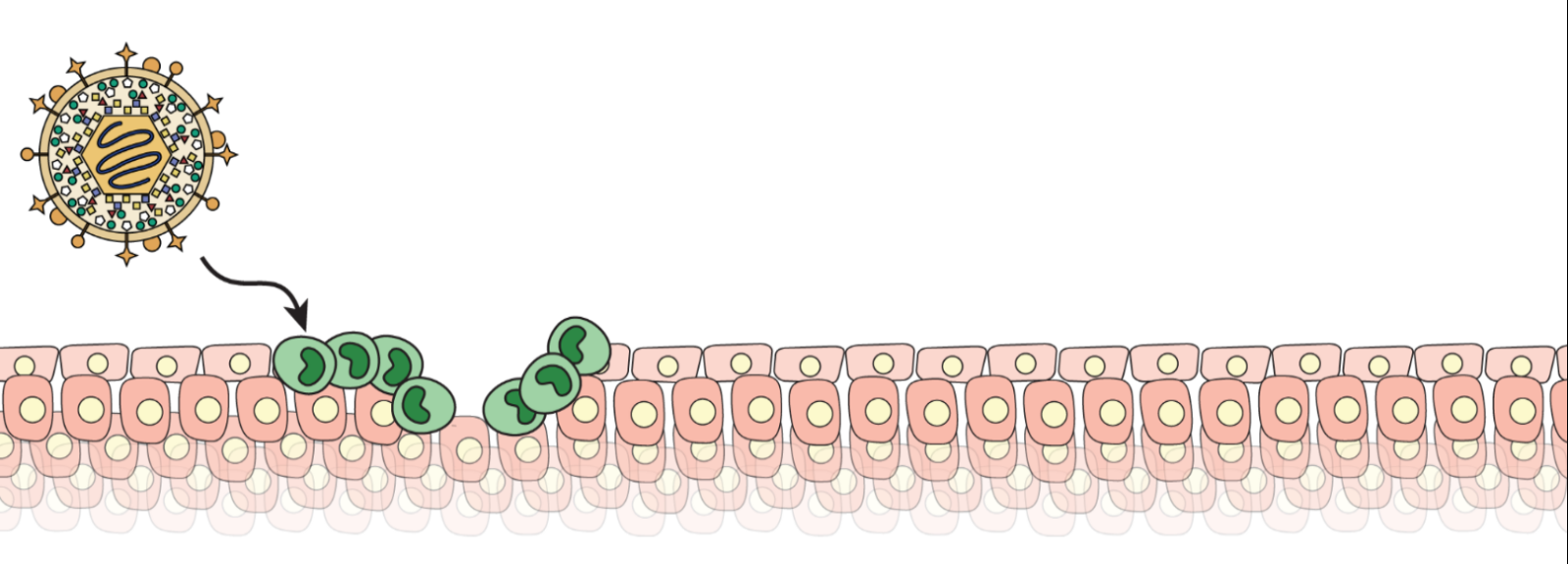
Human cytomegalovirus (HCMV) is a successful, widespread pathogen that infects the majority of the world’s population by early adulthood. The virus sustains a lifelong infection by establishing a reservoir of latently infected cells, as well as persistently infecting cells that intermittently shed infectious virus. The virus infects numerous cell types and can be found in most organ systems.
HCMV infection is a significant cause of morbidity and mortality in populations with immature or compromised immune systems. HCMV is the most common viral cause of birth defects, with congenital infection frequently resulting in hearing loss and developmental delays. HCMV infection contributes to graft loss in bone marrow and solid organ transplants and is the major cause of retinitis and blindness in AIDS patients. Even in individuals with competent immune systems, HCMV infection is associated with inflammatory and proliferative diseases such as certain cardiovascular diseases and some cancers.
Both primary infection and reactivation of latent infections cause HCMV disease. There is no vaccine to prevent HCMV infection, and the currently FDA-approved drugs for the treatment of HCMV disease suffer from low bioavailability, toxicity, and the development of viral resistance. Furthermore, existing antivirals are unable to eliminate latent reservoirs, underscoring the importance of better understanding this aspect of the viral life cycle in order to develop novel antiviral strategies.
How does the pathway of virus entry dictate the outcome of infection (lytic vs. latent)?
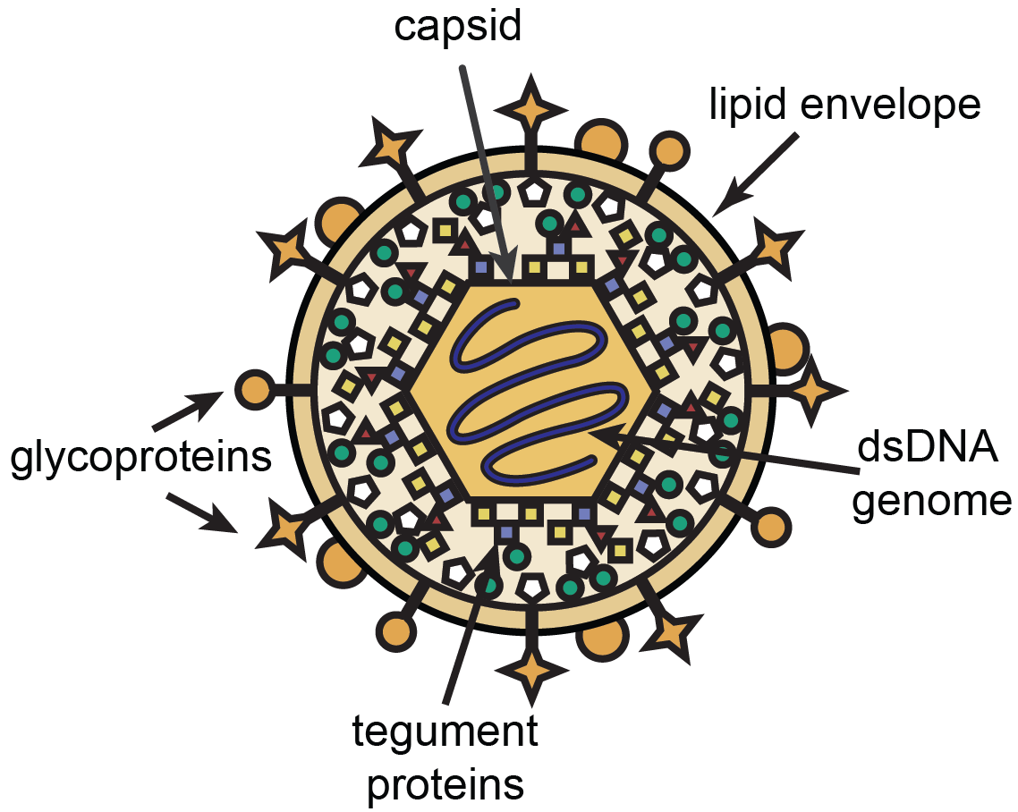
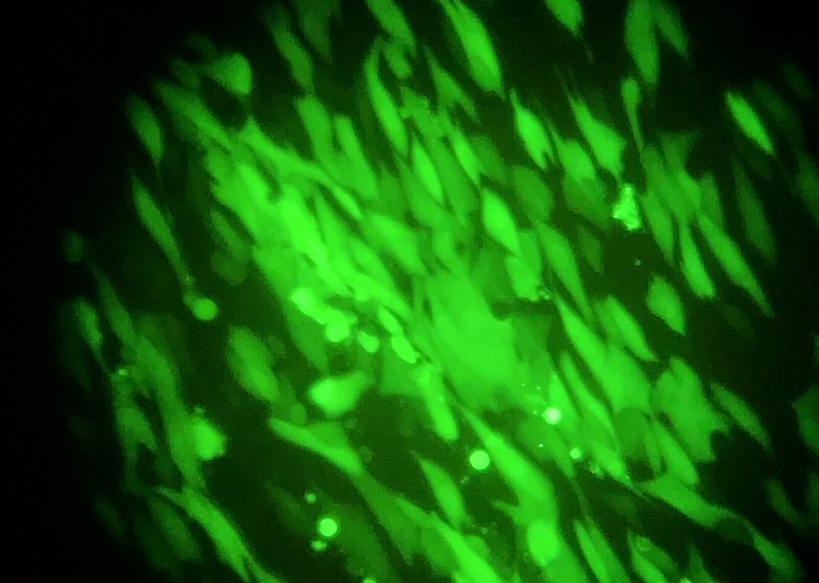
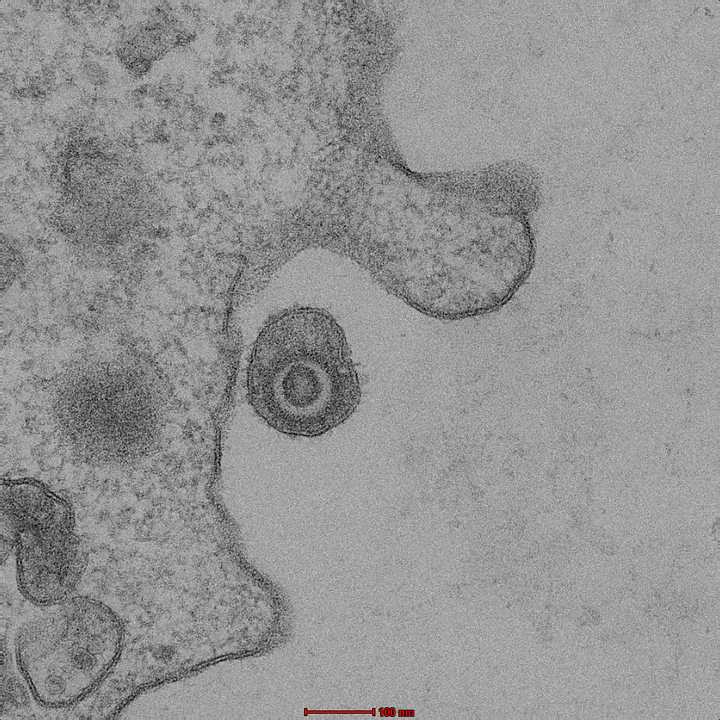
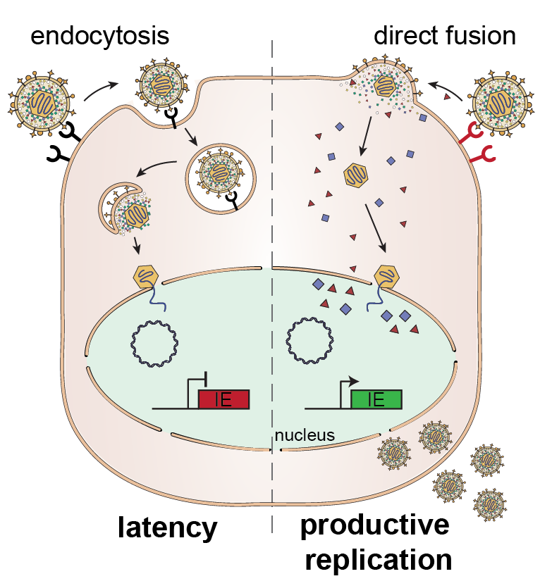
HCMV is an enveloped virus that can enter cells by either of two mechanisms. One is direct fusion of the virion envelope with the plasma membrane. The other is endocytosis of the intact virion into a cytoplasmic endosome and then subsequent fusion of the virion envelope and endosomal membrane. We have published that HCMV enters the incompletely differentiated myeloid cells in which it establishes latency by a form of endocytosis called macropinocytosis. During this entry process, viral proteins that are located within the virion tegument layer and important for turning on viral lytic genes remain associated with the endosome and are excluded from the nucleus. We showed that when HCMV enters these same cells by direct fusion, it delivers its tegument proteins to the nucleus and initiates a lytic infection. We are testing the hypothesis that the mechanism of entry dictates the subcellular localization of the tegument proteins and thus the infection outcome using sophisticated imaging techniques, differential receptor expression, and induced fusion approaches.
How is viral chromatin assembled?
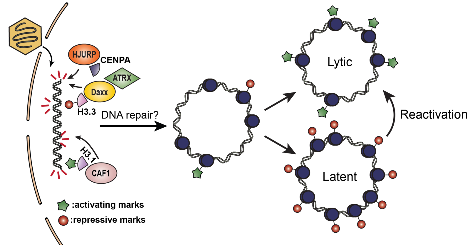
In the virion, the viral genome is naked but once it enters the nucleus, histones are rapidly deposited to create viral chromatin that is important for controlling viral gene expression. We are examining how this chromatin is initially assembled on incoming viral genomes and the impact it has on the outcome of infection.
When double stranded DNA is wrapped around histones, the DNA is more stable and the epigenetic modifications on the histones can amplify or diminish transcription. There are no histones associated with viral genomes inside herpesvirus virions, but once those viral genomes are injected into the nucleus, they are rapidly bound by histones. Histones are normally deposited onto cellular DNA during the process of DNA replication, transcription, or repair. We have shown that neither replication nor transcription is required for histone deposition onto infecting HCMV genomes, suggesting it may occur in response to DNA repair. Interestingly, herpesvirus virion genomes have linear double-stranded ends and contain nicks and gaps that represent repairable DNA damage. We are exploring the impact of cellular DNA repair pathways on the process of initial chromatinization of the HCMV genome using state of the art genome-wide histone mapping techniques and novel whole genome sequencing approaches.
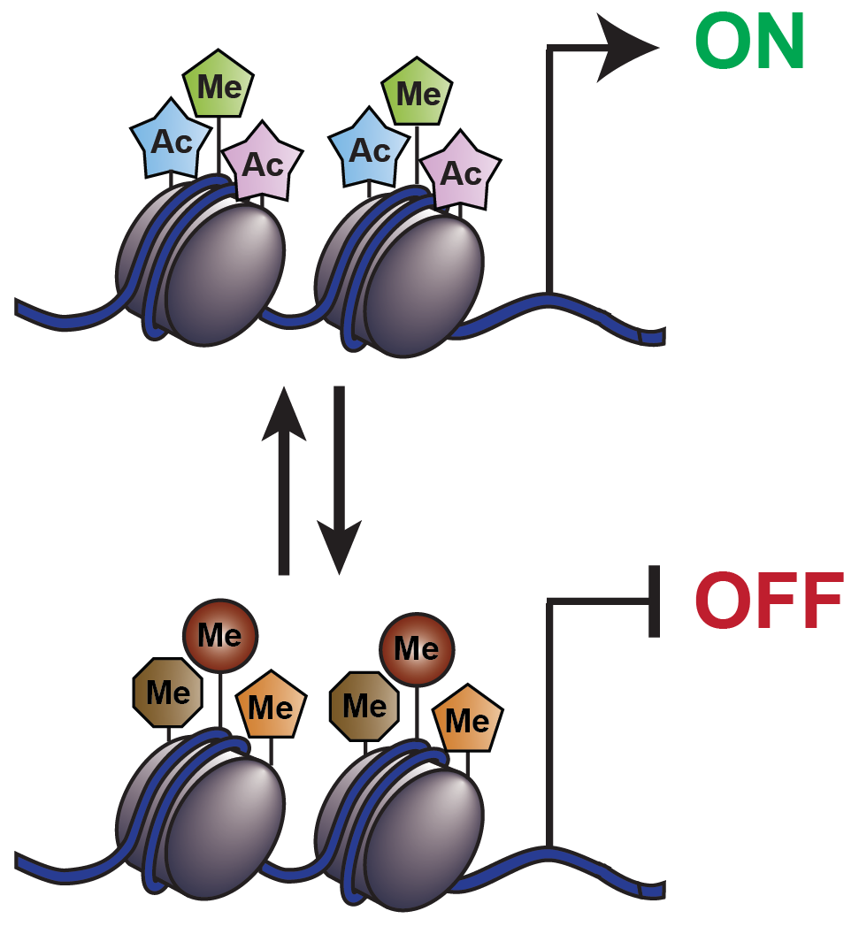
The modifications on histones associated with a gene dictate whether it will be transcriptionally active or repressed. We are defining how HCMV viral epigenetic structure is established and modified, and the impact on viral transcription and infection outcome.
Acetylated histones activate transcription and methylated histones repress transcription. We are examining whether the histones that get added to infecting HCMV genomes are modified prior to their deposition, and how their modifications change, in a cell type dependent manner, once they are associated with the viral genome. The ultimate goal is to understand how viral and cellular factors create and change the epigenetic signature of the viral genome and how that structure dictates viral transcription and infection outcome.
Cells have defenses against viruses, but HCMV has countermeasures. We explore the fascinating arms race between HCMV and the host for the ultimate control over the infected cell.
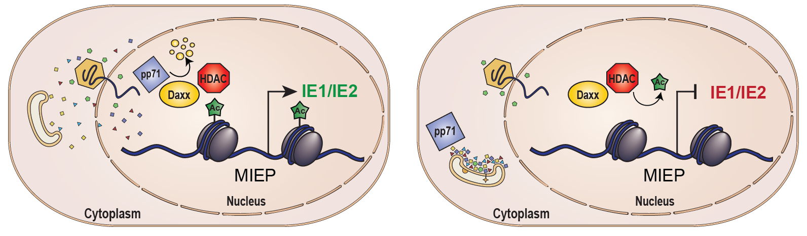
Intrinsic defenses are constitutively expressed and can act immediately against invading pathogens. A key intrinsic defense against HCMV is the cellular Daxx protein. We have shown Daxx assembles heterochromatin on the viral genome and silences viral transcription unless it is degraded by the viral tegument-delivered pp71 protein. We study how Daxx impacts the HCMV genome, and how pp71 migrates to the nucleus and induces Daxx degradation.
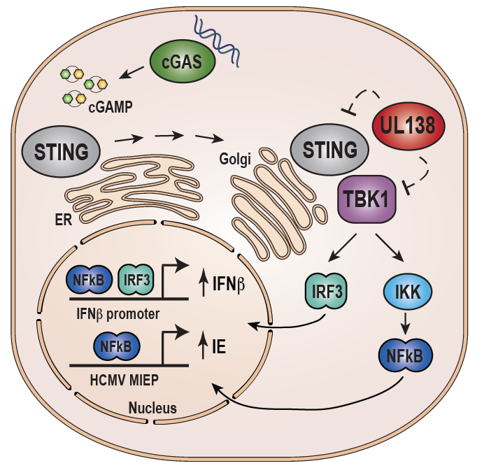
Innate defenses against HCMV include the cGAS/STING/TBK1 pathway. cGAS senses double stranded DNA in the cytoplasm and activates STING, which in turn activates the antiviral interferon response. Paradoxically, we have found that activation of this pathway also turns on viral gene expression during latency. We have identified a suite of HCMV latency genes, including the UL138 protein, that inactivate the STING pathway to enforce repression of viral lytic phase genes during latency. We study how UL138 modulates innate immunity and the role these proteins play during viral lytic and latent infections.
We study how HCMV modulates the division cycle of the infected cell and how the virus may contribute to human cancers like glioblastoma multiforme (GBM).
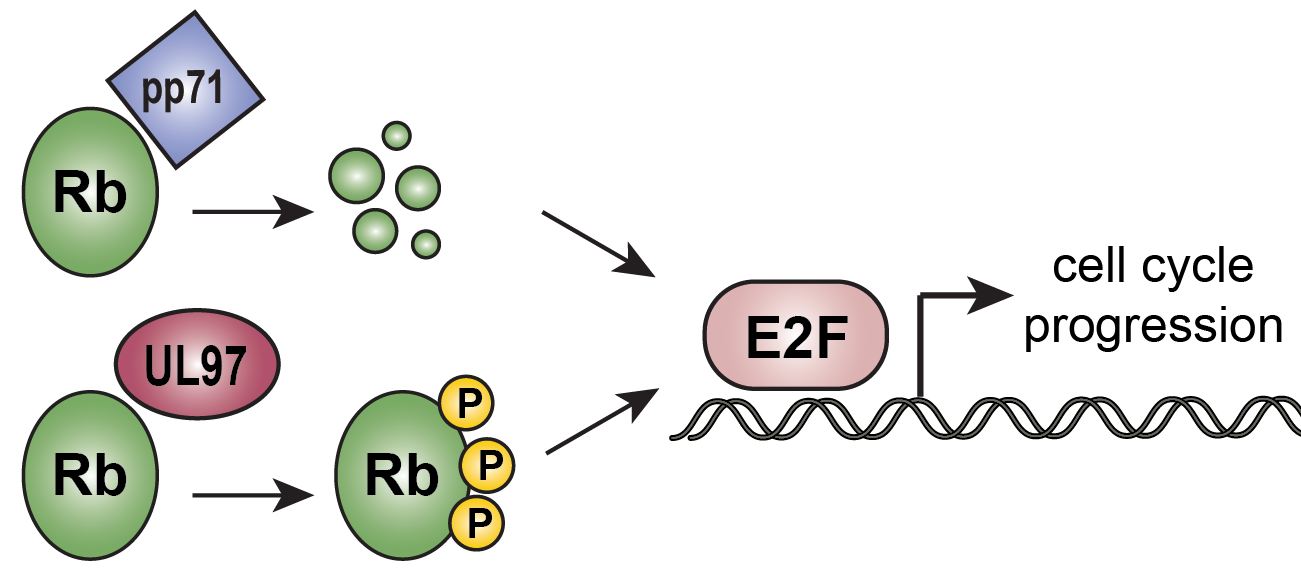
We have identified two HCMV proteins that inactivate the retinoblastoma (Rb) family of tumor suppressors to activate E2F-responsive gene expression and drive cell cycle progression. They are pp71 and UL97. HCMV pp71 binds to the three Rb family members (Rb, p107 and p130) and induces their proteasome-dependent, ubiquitin-independent degradation. UL97 is a kinase that phosphorylates all three Rb family members, disrupting their ability to bind and inhibit the cellular E2F transcription factors that are necessary for cell cycle progression. We continue to study the impacts of Rb inactivation by pp71 and UL97 on HCMV infection and oncogenesis, and to identify additional substrates for both HCMV proteins.
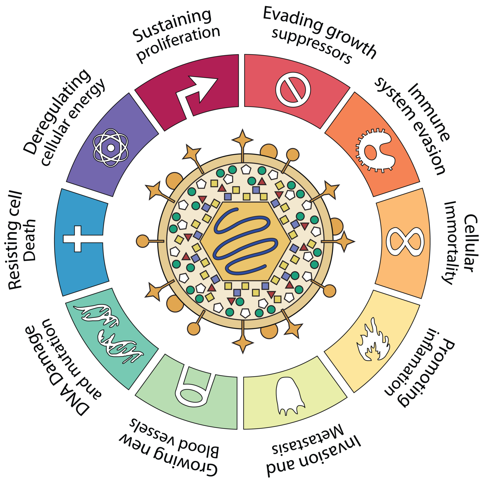
HCMV infection is associated with certain types of cancer including gliobastoma (GBM). Unlike other tumor viruses, HCMV infection is not known to directly transform cells. However, HCMV has to capacity to promote all of the hallmarks of cancer.
Interestingly, while we find HCMV genomes present in GBM tumor samples, not all cells within the tumor harbor viral genomes, suggesting that, if HCMV is driving the cancer, it must do so in a way that is distinct from other tumor viruses. We found HCMV infection of GBM cells increases their resistance to the chemotherapeutic drug Temozolomide and their ability to self-renew, making them become more like a cancer stem cell. Thus we hypothesize that rather than directly transforming cells, HCMV infection may protect a small fraction of tumor cells from chemotherapy-induced cell death, and thereby accelerate tumor regrowth after therapy withdrawal.